
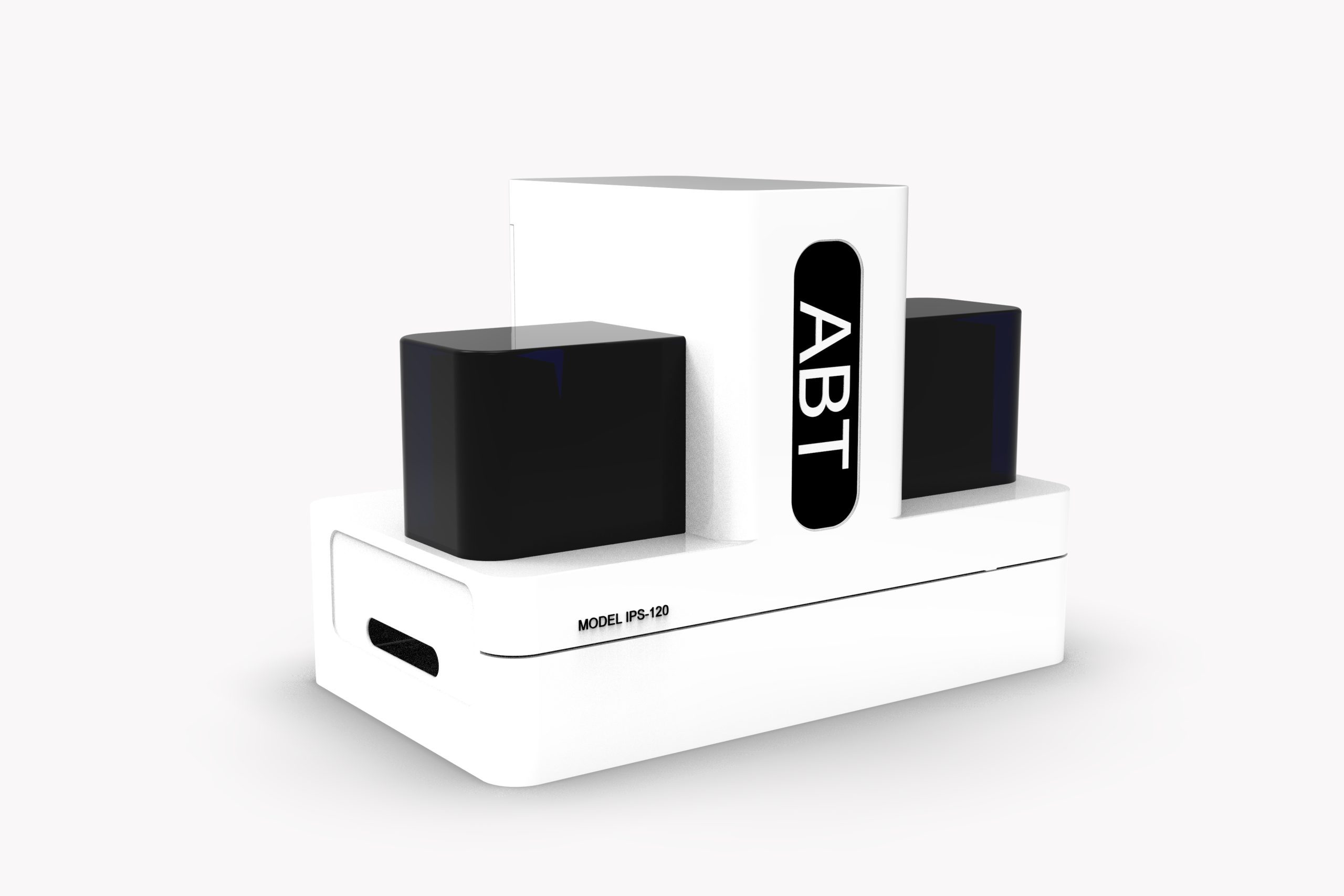

INTRODUCTION TO INNOCELL PATHOLOGY SCANNER
- InnoCell Pathology Scanner (IPS-120) , also known as a digital slide scanner, utilizes advanced Whole Slide Imaging (WSI) technology and is engineered for the high-throughput scanning of up to 120 slides simultaneously. The device features full automation of slide transport, real-time focusing, and seamless image stitching to produce high-resolution digital slides. The IPS-120 addresses the rigorous demands of cytological diagnosis and research, facilitating the standardization, digitization, and digital transformation (Digital Cytology) of clinical workflows.
- System Configuration: Optical system, image acquisition system, scanning software, slide chamber, and control module.
- Intended use: The IPS-120 is designed for the scanning, digitization, and visualization of cytological specimens to support:
- Scanning and converting traditional cytological slides into high-definition digital images for diagnostic purposes and morphological evaluation.
- Providing comprehensive datasets and high-quality imagery to facilitate advanced research and academic training in cytology.
- Automating the entire scanning procedure to minimize manual intervention and human error.
- Enabling the secure storage, rapid retrieval, and seamless sharing of digital slide data.
- Supporting the integration of digitized and standardized workflows within laboratory environments.
- The device is specifically optimized for:
- Hospitals and diagnostic centers
- Laboratories and clinics
- Biomedical research and academic institutions
- Organizations committed to digital transformation

OPERATION PRINCIPLE
InnoCell Pathology Scanner (IPS-120) operates on an automated slide-loading mechanism integrated with high-precision microscopy and digital imaging systems. During the scanning process, the device performs real-time autofocusing while capturing continuous fields of view. The integrated software seamlessly stitches these images together to create a high-definition Whole Slide Image (WSI), enabling comprehensive specimen analysis directly on a computer monitor.
- Automated Slide Loading
Slides are housed in a high-capacity loading system that holds up to 120 specimens (30 trays, 4 slides per tray). The system automatically identifies and processes slides in sequence for maximum workflow efficiency. - Intelligent Real-Time Scanning & Autofocus
Utilizing high-magnification objectives combined with a real-time autofocus mechanism, the IPS-120 ensures crisp, sharp imaging across the entire scanned area without the need for manual intervention. - Seamless Image Stitching
Individual image fields are processed and stitched by advanced software to create a unified digital slide, ensuring total consistency in brightness, color balance, and resolution. - Data Export & Archiving
Digitized slides are immediately available for viewing, analysis, and archiving. The system supports easy integration into Laboratory Information Systems (LIS) and other digital pathology workflows.
KEY FEATURES
- High Throughput: Batch scanning capacity of up to 120 cytological slides.
- Rapid Scanning Speed: Less than 45 seconds per slide (with high-speed modes reaching 18 seconds).
- Superior Image Quality: Equipped with a 20X APO / 0.8 NA objective and a resolution of 0.25 µm/pixel, meeting the stringent requirements for detailed morphological analysis in clinical diagnostics and cytological research.
- User-Friendly Software & Operation:
- Automated Barcode Recognition for streamlined patient record management.
- Real-time monitoring of the scanning process.
- Queue Management: Preview and manage lists of slides currently being scanned or pending in the queue.
- Multi-tasking Capability: Simultaneous processing of 4 slides at once.
SPECIFICATIONS
Model | IPS-120 |
| Sample Capacity | 120 slides (Standard Slide Size: 25 mm x 75 mm) |
| Scanning Speed | < 45 seconds (15 mm × 15 mm area, 20X objective); as fast as 18 seconds |
| Scanning Camera | 5.0MP Color CMOS |
| Preview Camera | 8.0 MP |
| Objective Lens | Olympus 20X/ 0.8 NA (Standard) Olympus 40X/ 0.95 NA (Optional) |
| Pixel Resolution | 0.25 µm/pixel (20X) |
| Focus Mode | Real-time Autofocus |
| Movement Stage | Motorized XYZ Stage |
| Image Formats | JPEG / JP2000 / TIFF / BMP / SVS / SCN |
| Light Source | Light Source |
| Dimensions (W×D×H) | 76 cm × 45 cm × 55 cm |
| Weight | ~43 kg |
Read more: InnoCell Pathology Scanner User Manual
Other products: DPIS – Digital Pathology Information System


Reviews
There are no reviews yet.