

About ABT ® GRAM STAINING KIT
ABT ® Gram Staining Kit is designed for staining and differentiating Gram-positive bacteria (purple to dark violet) and Gram-negative bacteria (pink to red) in cultured bacterial samples and clinical specimens. It is intended for use in diagnostic microbiology to support infection diagnosis and in microbiological research.
Sample types: Cultured bacterial isolates or clinical specimens
Staining characteristics: Bacterial cells stained with the kit exhibit a distinct contrast between Gram-positive bacteria (purple to dark violet) and Gram-negative bacteria (pink to red).
Detection capability: Cultured bacterial samples and clinical specimens processed and stained with the kit allow identification of a wide range of Gram-positive and Gram-negative bacteria of varying shapes and sizes. In addition, the kit facilitates visualization of fungal elements in clinical specimens following staining.
Principle of operation:
- Based on differences in the peptidoglycan layer between Gram-positive and Gram-negative bacteria, which determines their ability to retain crystal violet dye.
- The staining procedure of ABT ® Gram Staining Kit consists of four main steps:
- First, fixed bacterial cells are stained with Crystal violet Solution, in which dye crystals bind to negatively charged proteins within the peptidoglycan layer of the bacterial cell wall.
- Next, the cells are treated with Lugol Solution, forming a Crystal Violet–Iodine (CV-I) complex that stabilizes and locks the crystal violet dye within the peptidoglycan.
- Subsequently, Gram differentiation is achieved using Decolorizer Solution, a mixture of alcohol and acetone that removes the outer membrane of Gram-negative bacteria and washes out the CV-I complex. In Gram-positive bacteria, the thick peptidoglycan layer retains the CV-I complex, resulting in a purple to dark violet coloration.
- Finally, the cells are counterstained with Safranin O Solution, a positively charged dye that binds to negatively charged cytoplasmic proteins, thereby visualizing Gram-negative bacteria in pink to red after staining.
Expiration date: 12 months from manufacture
Storage condition: Nhiệt độ 15 – 30℃, tránh sáng trực tiếp.
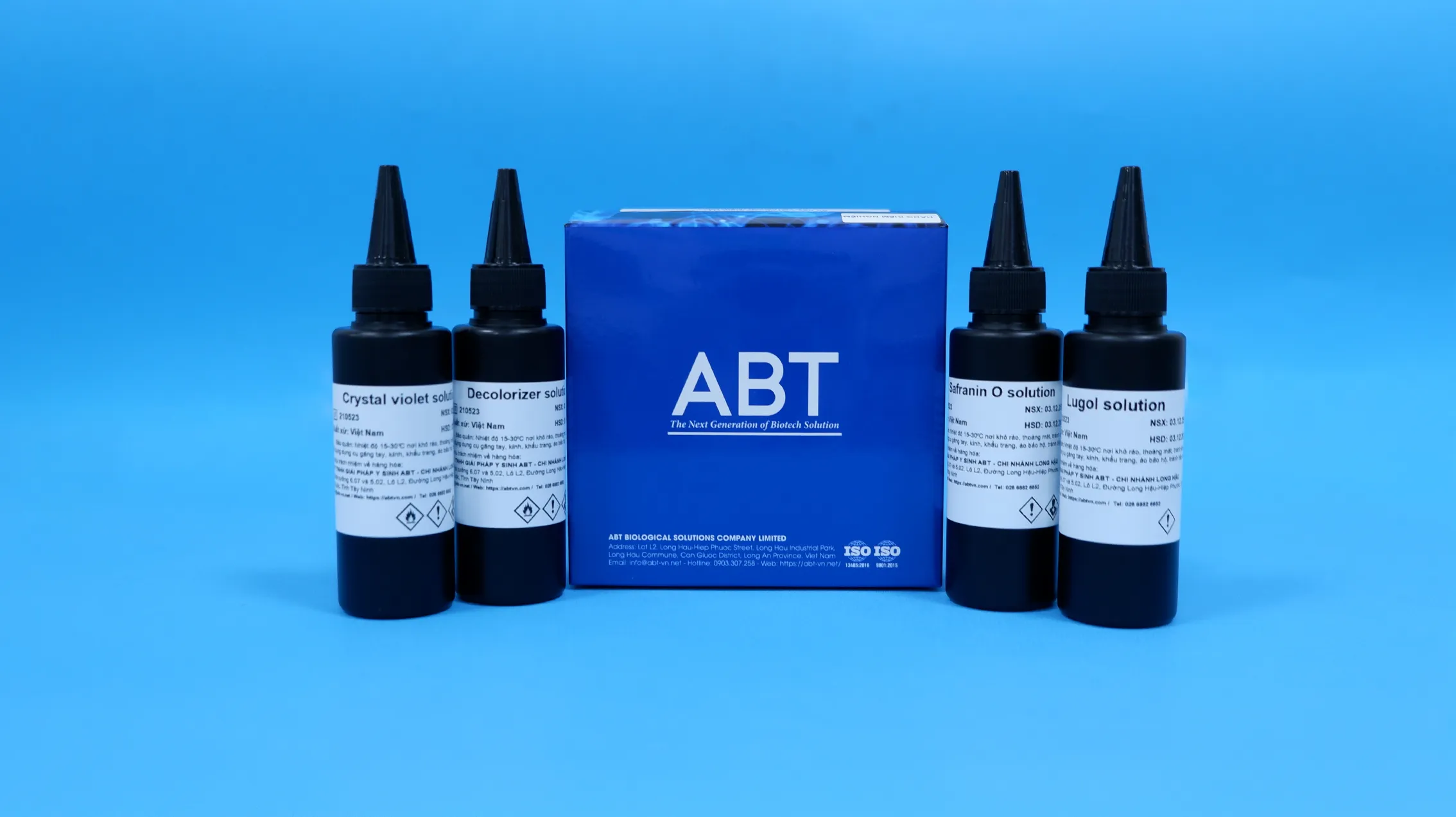
Components:
| No. | KIT COMPONENTS | QUANTITY |
| 1 | Crystal Violet Solution | 1×100 mL |
| 2 | Lugol Solution | 1×100 mL |
| 3 | Decolorizer Solution | 1×100 mL |
| 4 | Safranin O Solution | 1×100 mL |
Procedure
1. Preparation of smears:
- Slide preparation: Immerse slides in 96% ethanol, allow to drain, and briefly flame over an alcohol lamp to clean. Label specimen information on the frosted end of the slide.
- Smear preparation: For cultured bacteria (colonies or liquid cultures) and clinical specimens (body fluids, urine, sputum, swabs, tissue), pre-process and homogenize according to national guidelines (Vietnam Ministry of Health Decision No. 1539/QĐ-BYT) or relevant veterinary/aquaculture/research regulations.
Smear preparation procedure:
- Step 1: Pipette 5–10 µL of processed sample directly onto the slide (surface swabs may be applied directly).
- Step 2: Spread the sample evenly in a spiral pattern from the center outward (urine samples may be applied without spreading).
- Step 3: Fix the smear by air-drying, then gently flame the underside of the slide 2–3 times (heat fixation), or immerse in 99.5% methanol for 1 minute (alcohol fixation).
Note:
- Smears must be evenly spread; overly thick or thin smears affect staining quality.
- Samples should be processed and fixed within 24–48 hours of collection to prevent bacterial degradation.
- Heat fixation must be performed quickly to avoid damaging Gram layers or distorting bacterial morphology.
2. Gram Staining Procedure
– Perform Gram staining of the prepared smear according to the following procedure:
- Step 1: Stain with Crystal Violet Solution for 30 seconds; rinse with tap water.
- Step 2: Fix with Lugol Solution for 30 seconds; rinse with tap water.
- Step 3: Decolorize with Decolorizer Solution for 30 seconds; rinse with tap water.
- Step 4: Counterstain with Safranin O Solution for 1 minute; rinse with tap water.
- Step 5: Allow slides to air dry.
Note:
- Apply 0.5–1.0 mL of reagent or sufficient volume to fully cover the smear.
- Tap water used for rinsing should have pH 7.0–7.5; if outside this range, use double-distilled water.
- Avoid direct mechanical impact during reagent application and rinsing to prevent detachment of samples, especially Gram-negative bacteria.
3. Result analysis
Analyze the results under a light microscope. Observe with the 10× and 40× objectives to assess the staining and decolorization quality of the smear. Examine with the 100× oil immersion objective to evaluate bacterial morphology, size, and Gram reaction: Gram-positive bacteria appear purple to dark violet, while Gram-negative bacteria appear pink to red.
Read more: ABT Bio-reagents ; Instructional Video for ABT Kit


Reviews
There are no reviews yet.